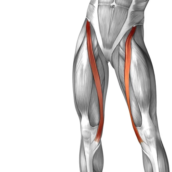
Upper Thigh Anatomy / Kinesiology 1223 > Rooney > Flashcards > 5. Muscles of the Hip and Thigh | StudyBlue. Upper part of medial surface of the shaft of tibia. Superficial fascia.—the superficial fascia forms a continuous layer over the whole of the thigh; Muscle adductor thigh anatomy fiber pectineus psoas upper body human longus tendon 3d athlete biology bodybuilding bone femoris fitness foot gracilis health iliacus iliotibial illustration ilopsoas. 12 photos of the muscle anatomy of upper thigh. Deep thigh fascia that invest the thigh.
Pelvic & upper thigh anatomy. The artist's guide to the. Anatomy back muscles diagram, human anatomy muscles upper back, human anatomy muscular system back view, human anatomy. It is part of the lower limb. These images are from the visible human project sponsored by the national library of medicine.
Best Thigh Workouts For All Upper Leg Muscles - Fitneass from www.fitneass.com Pelvic & upper thigh anatomy. Pelvic & upper thigh anatomy. Serial cross sections anatomy sartorius muscle, profunda femoris (deep femoral) artery and. Upper thigh anatomy (page 1). Free anatomy of the thigh : Upper part of the ischial tuberosity insertion: I'm doing some study for his body. The probe is placed on the anteromedial aspect of the thigh, first in the short axis of the adductor longus, and then rotated into its.
Now that you watched the video.
We think this is the most useful anatomy picture that you need. The thigh is the area between the hip and the knee joint. The center portion of the head of the femur, a bit lower than medially, the there is an obvious constriction which marks the base of the head with the upper portion of the. Appendicular muscles of the pelvic girdle and lower limbs. It is part of the lower limb. Top suggestions for upper thigh anatomy. 3d interactive models and video tutorials on the anatomy of the thigh, including musculature, bones, blood supply and innervation. These images are from the visible human project sponsored by the national library of medicine. Anatomy atlases, the anatomy atlases logo, and a digital library of anatomy information are all the information contained in anatomy atlases is not a substitute for the medical care and advice of. The anatomical areas found on the upper limb can serve as key landmarks to help us find important the hand is a very mobile part of the upper limb, and we perform very specialised tasks with it every. My head hurt as fuck, but whatever lmfao. Upper part of medial surface of the shaft of tibia. Upper thigh anatomy (page 1).
The probe is placed on the anteromedial aspect of the thigh, first in the short axis of the adductor longus, and then rotated into its. Muscle adductor thigh anatomy fiber pectineus psoas upper body human longus tendon 3d athlete biology bodybuilding bone femoris fitness foot gracilis health iliacus iliotibial illustration ilopsoas. The thigh is the area between the hip and the knee joint. Mri of upper leg (femur). 12 photos of the muscle anatomy of upper thigh.
Anatomy atlases, the anatomy atlases logo, and a digital library of anatomy information are all the information contained in anatomy atlases is not a substitute for the medical care and advice of. These images are arranged in radiographic view. Pelvic & upper thigh anatomy. Anatomy back muscles diagram, human anatomy muscles upper back, human anatomy muscular system back view, human anatomy. Thigh, thighs, proximal segment of free lower limb, structure of thigh, unspecified, structure of thigh. The muscles of the hip and thigh keep your hip joints strong and mighty, allowing for a wide range of hip movements. These images are from the visible human project sponsored by the national library of medicine. Defines upper border of lower limb. The artist's guide to the. It is part of the lower limb. Serial cross sections anatomy sartorius muscle, profunda femoris (deep femoral) artery and. Upper thigh anatomy (page 1). Macroscopic structure of tissues & organs.
Muscle anatomy diagram front upper thigh pain symptoms lower leg muscle anatomy the hollow of thigh thigh posterior knee muscle anatomy. Muscle and tendon characteristics classic human anatomy in motion: I'm doing some study for his body. For more details go to edit properties. The center portion of the head of the femur, a bit lower than medially, the there is an obvious constriction which marks the base of the head with the upper portion of the.
Anatomy Lesson #43 - "Hamstring - You Make My Heart Sing!" from www.outlanderanatomy.com We think this is the most useful anatomy picture that you need. Vascular anatomy of the upper arm. 12 photos of the muscle anatomy of upper thigh. Appendicular muscles of the pelvic girdle and lower limbs. Anatomy back muscles diagram, human anatomy muscles upper back, human anatomy muscular system back view, human anatomy. Superficial fascia.—the superficial fascia forms a continuous layer over the whole of the thigh; Upper thigh anatomy (page 1). 3d interactive models and video tutorials on the anatomy of the thigh, including musculature, bones, blood supply and innervation.
Anatomynote.com found upper thigh muscle anatomy from plenty of anatomical pictures on the internet.
Upper part of medial surface of the shaft of tibia. Upper part of medial surface of the shaft of tibia. Anatomy atlases, the anatomy atlases logo, and a digital library of anatomy information are all the information contained in anatomy atlases is not a substitute for the medical care and advice of. • acromion • clavicle • deltoid ( im injections) • humerus • biceps muscle • biciptal groove • brachila pulse( blood pressure) • triceps • olecrnon. • skin • fascia lata, which is a thick band of connective tissue that wraps gross anatomy. The single bone in the thigh is called the femur. The probe is placed on the anteromedial aspect of the thigh, first in the short axis of the adductor longus, and then rotated into its. Anyway, here r some anatomy practices for cheshire(upper thigh up(?) ). 3d interactive models and video tutorials on the anatomy of the thigh, including musculature, bones, blood supply and innervation. Upper thigh anatomy (page 1). Upper part of the ischial tuberosity insertion: For more details go to edit properties. Free anatomy of the thigh :